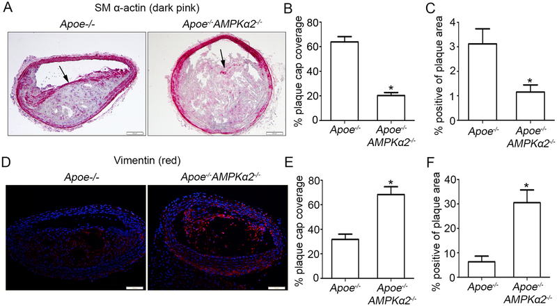
Figure 2. AMPKα2 deletion induces VSMC phenotypic switching in advanced atherosclerotic plaque in the BA.
(A) Representative images of IHC staining of SM α-actin (dark pink) in BA of Apoe−/− and Apoe−/−AMPKα2−/− mice. Arrow represents representative staining of SM α-actin. Scale bar=100 μm. (B) Quantification of plaque SM α-actin coverage on the plaque cap in BA of Apoe−/− and Apoe−/−AMPKα2−/− mice. (C) Quantification of total plaque SM α-actin content in BA of Apoe−/− and Apoe−/−AMPKα2−/− mice. (D) Representative images of IF staining of vimentin (red) in BA of Apoe−/− and Apoe−/−AMPKα2−/− mice. Dapi = blue staining of nucleus. Scale bar=100 μm. (E) Quantification of plaque vimentin coverage on the plaque cap in BA of Apoe−/− and Apoe−/−AMPKα2−/− mice. (F) Quantification of total plaque vimentin content in BA of Apoe−/− and Apoe−/−AMPKα2−/− mice. n=10 in each group. Values represent the mean ± SEM. *, P<0.05 vs. Apoe−/− mice.

