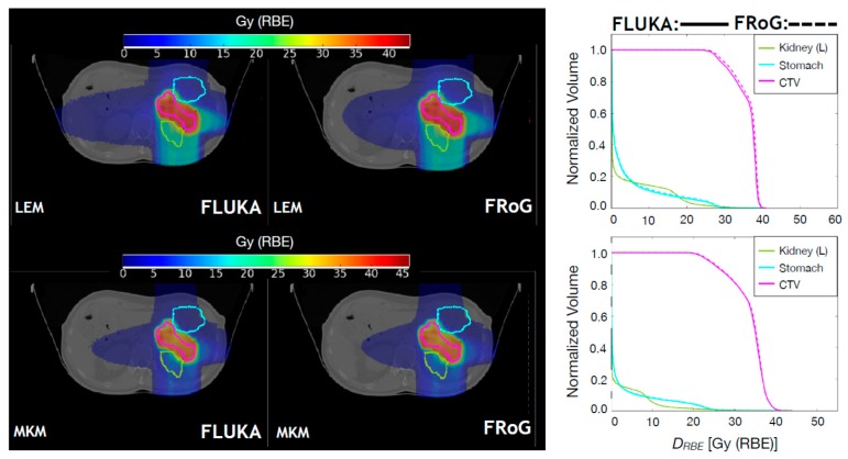
Figure 5.
Upper panels: FLUKA and FRoG recalculated LEM-based DRBE distributions for a 12C ion pancreatic patient case are displayed in the left and middle panel, respectively, together with the contours of three representative regions of interest: CTV, left kidney and stomach. The LEM-based DRBEVH for the ROIs are shown in the right panel. Lower panels: FLUKA and FRoG recalculated MKM-based DRBE distributions for a 12C ion pancreatic patient case are displayed in the left and middle panel, respectively, together with the contours of three representative regions of interest: CTV, left kidney, and stomach. The MKM-based DRBEVH for the ROIs are shown in the right panel.

