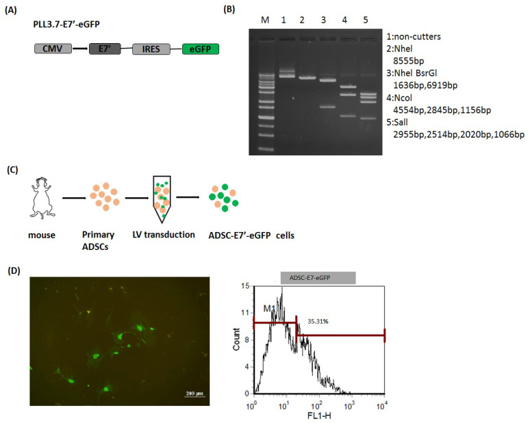
Figure 1.
Establishment of ADSC labeled with enhanced green fluorescent protein (eGFP) and combined with modified E7’ (ADSC-E7’-eGFP). (A) Schematic diagram of pLL3.7-E7’-eGFP construction; (B) agarose gel electrophoresis of plasmid pLL3.7-E7’-eGFP (M: 1 kb DNA ladder; lane 1: Undigested plasmid; lane 2: uNheI (8555 bp); lane 3: NheI and BsrGI (1636 bp and 6919 bp); lane 4: bNcoI (4554 bp, 2845 bp, and 1156 bp); lane 5: bSalI (2955 bp, 2514 bp, 2020 bp, and 1066 bp)); (C) illustration of lentiviral transduction of primary ADSCs; and (D) fluorescence microscopy and flow cytometric analysis of ADSC-E7’-eGFP cells.

