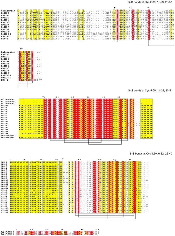
Figure 3.
Multiple sequence alignment of the KVs toxins in A. viridis. Alignment was performed with the T-coffee tool [43]. Similar residues are written in bold characters and boxed in yellow, whereas conserved residues are in white bold characters and boxed in red. The sequence numbering on the top refers to the alignment. For each alignment, the pattern of Cys residues forming disulfide bridges is shown. Type 1, 2, 3 and 5 KV blockers are reported; while no member of Type 4 has been identified to date. No S-S bonds and Cys pattern are defined for type V KTx because of the absence of any 3D structure experimentally determined to date.

