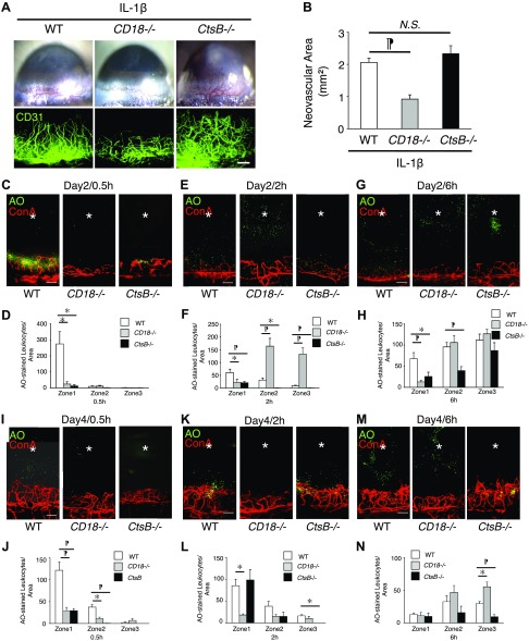
Figure 4.
Ex vivo leukocyte transmigration rate during IL-1β-induced angiogenesis in CD18−/− and CtsB−/− mice. To illustrate a large region of the corneal flatmount at higher resolution, composite micrographs were generated by merging the digital images from adjacent regions of the cornea in a mosaic fashion. A) Microscopic pictures of IL-1β-induced corneal neovascularization of WT, CD18−/−, and CtsB−/− mice. Double staining of corneal flatmounts for angiogenesis (CD31). B) Quantitative analysis of vascular area (n = 8–9). The neovascular area was determined by performing CD31 immunohistochemistry and measuring the vascular area. C–H) AO+ leukocytes and Con A+ angiogenic vessels in IL-1β-implanted corneas of CD18−/− and CtsB−/− mice at 0.5 (C), 2 (E), and 6 (G) h after AO injection on d 2 after pellet implantation. Quantitation of the number of AO+ leukocytes in IL-1β-implanted corneas at 0.5 (D), 2 (F) and 6 (H) h after AO injection on d 2. I–N) AO+ leukocytes and Con A+ angiogenic vessels in IL-1β-implanted corneas of CD18−/− and CtsB−/− mice at 0.5 (I), 2 (K) and 6 (M) h after AO injection on d 4 after the pellet implantation. Quantitation of the number of AO+ leukocytes in IL-1β-implanted corneas at 0.5 (J), 2 (L) and 6 (N) h after AO injection on d 4 (n = 4). *P < 0.05, ¶P < 0.01. Scale bars, 100 µm.

