Figure 6.
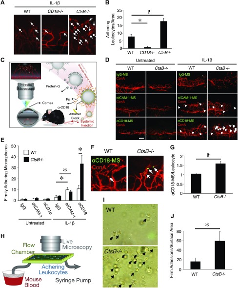
Leukocyte adhesion in IL-1β-induced angiogenesis of CtsB−/− mice. To illustrate a large region of the corneal flatmount at higher resolution, composite micrographs were generated by merging the digital images from adjacent regions of the cornea in a mosaic fashion. A) Representative micrographs of flatmounted IL-1β-implanted corneas of WT, CD18−/−, and CtsB−/− mice at 4 d after pellet implantation. Firmly adherent leukocytes in the corneal neovasculature were visualized by perfusion with ConA. Arrows: firmly adherent leukocytes in the inflamed corneal neovasculature. B) The average number of firmly adherent leukocytes in the inflamed corneal neovasculature of WT, CD18−/−, and CtsB−/− mice (n = 4–6). WT, 7.8 ± 1.5/area; CD18−/−, 0.8 ± 0.5; and CtsB−/−, 17.8 ± 2.1. C) Experimental design of our in vivo molecular imaging assay. D) α-IgG, α-ICAM-1 mAb or α-CD18 mAb–conjugated microspheres (MSs) (green) and rhodamine-conjugated ConA (red). Arrows: firmly adherent MSs in blood vessels. E) Quantitation of the number of MSs in corneal vessels of untreated and IL-1β-implanted eyes (d 4; n = 4–7). F) Ex vivo visualization of accumulated MSs in corneal vessels: α-CD18 mAb–conjugated MSs and rhodamine-conjugated ConA. Arrows: adherent MSs. G) Quantification of the firmly adherent MSs in corneal vessels. H) The design of the microfluidic mouse leukocyte adhesion experiments. I) Representative micrographs of firmly adherent leukocytes (arrows) from WT and CtsB−/− mice. J) Quantitation of the number of firmly adherent leukocytes in WT and CtsB−/− mice at a shear stress of 2.5 dyn/cm2. *P < 0.05, ¶P < 0.01. Scale bars, 100 µm.

