Figure 7.
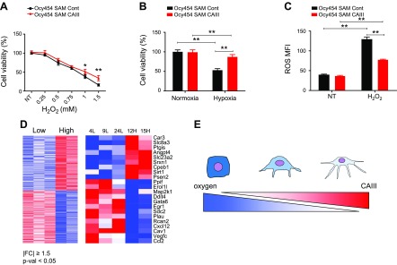
CAIII overexpression protects osteocytes from cell death and ROS. A) CAIII-overexpressing Ocy454 cells (SAM-CAIII) displayed an increase in cell viability and reduced sensitivity to H2O2 exposure at various doses. B) SAM-CAIII cells were exposed to hypoxia (1% O2) for 24 h. C) H2O2-induced ROS activity is reduced in SAM-CAIII cells compared with SAM control. D) Unbiased global transcriptome analysis with microarray (Affymetrix) identified 518 genes (left) that were significantly up- or down-regulated in high-Sost–expressing clones (|FC| ≥1.5; P < 0.05; Supplemental Table 1). Expression values are shown as a colored representation (heatmap) in which each row corresponds to an Entrez Gene ID and each column corresponds to a clone (from left to right: 4L, 9L, 24L, 12H, and 15H). Red and blue indicate expression values ≥2 sd above and below, respectively, the row-wise mean (white) computed across all clones. Among those 518 genes, Functional Annotation Tools (DAVID) indicated that 22 genes (right), including CAIII, were involved in the response to hypoxic and/or oxidative stress (Supplemental Table 2). E) Schematic diagram of the relationship between CAIII expression and oxygen levels in osteocytic cell differentiation. As osteoblasts differentiate into osteocytes after being entrapped in the mineralized bone matrix in vivo, the oxygen level available for differentiating osteoblasts is decreased, whereas the expression of CAIII, which protects osteocytes from cell death and ROS, is increased.

