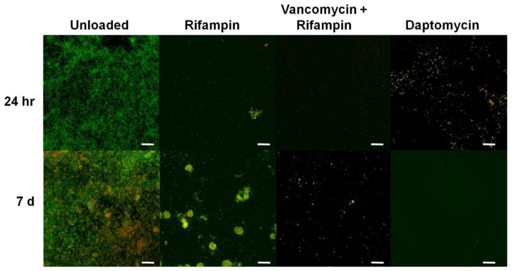
Figure 4.
Representative CSLM images showing S. epidermidis ATCC 35984 biofilm (live cells stained green and dead and membrane compromised cells stained red or yellow) following treatment for 24 h and 1 week with unloaded beads (negative control) and beads loaded with rifampicin (Rifampin), rifampicin and vancomycin, and daptomycin. Scale bars: 25 µm.

