Figure 1.

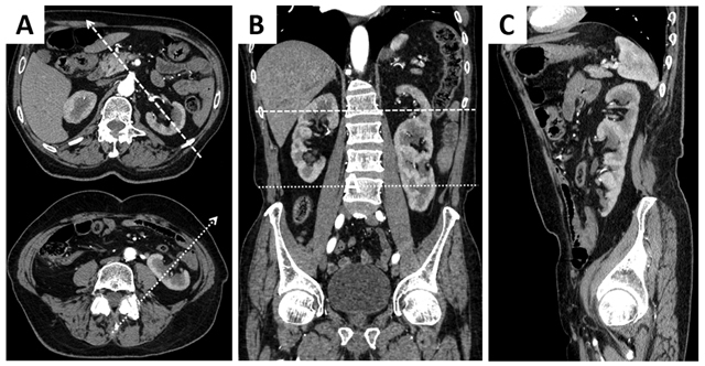
Contrast-enhanced CT with axial (A), coronal (B) en sagittal (C) reformations demonstrating a large, S-shaped kidney in the left renal fossa and a normal kidney on the right.

Contrast-enhanced CT with axial (A), coronal (B) en sagittal (C) reformations demonstrating a large, S-shaped kidney in the left renal fossa and a normal kidney on the right.