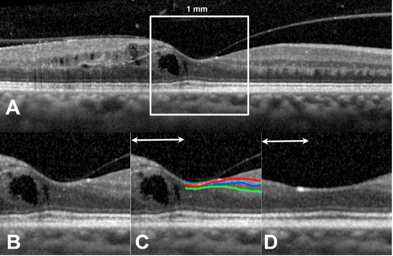
Figure 1. Representative optical coherence tomography images of a study patient.
Disorganization of retinal inner layers (DRIL) was evaluated in the central 1 mm of the line scan centered on the fovea (A). This 1 mm-wide portion (B) was evaluated for the boundaries between the ganglion cell layer-inner plexiform layer (red line), inner plexiform layer-inner nuclear layer (blue line), and inner nuclear layer-outer plexiform layer (green line). The horizontal extent of disruption of for which boundaries between any of these layers could not be identified was measured as DRIL (line with arrowheads). (C) An area of DRIL overlying a cyst of intraretinal fluid. Thirty months later, the fluid had resolved (D) but DRIL was still present.

