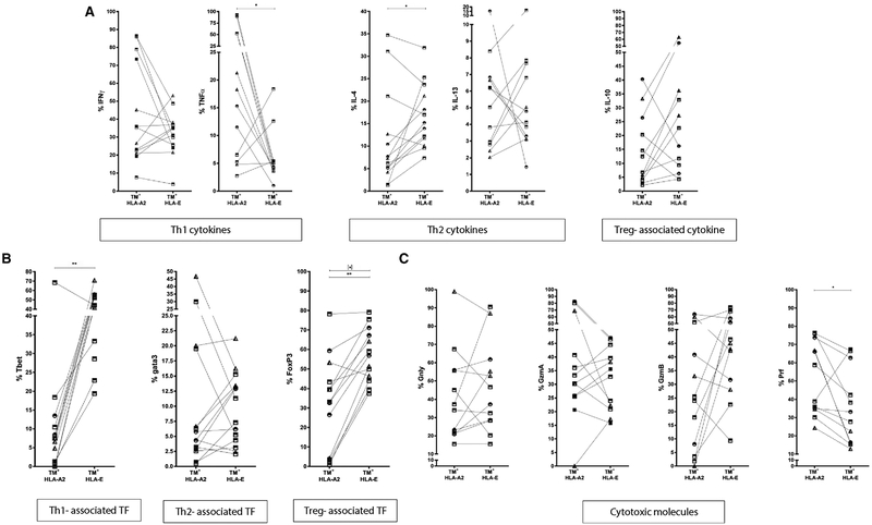
Figure 5.
Comparison of HLA-A*0201 and HLA-E TM+ CD8+ T cells. PBMCs were expanded using mitogenic stimulation, followed by magnetic bead separation of CD8+ T cells and specific peptide stimulation. Cells were stained with TMs first before staining with cell surface markers and intracellular cytokine staining. Frequency of HLA-A*0201/Mtb and HLA-E/Mtb TM+ CD8+ T cells producing IFNγ, TNFα, IL-4, IL-13, and IL-10 (A), expressing T-bet, Gata3 and FoxP3 transcription factors (B) and producing Granulysin, Granzyme A, Granzyme B and Perforin (C). 3 out 26 HDs (all Dutch), 3 out 13 LTBI, 7 out 17 active TB patients for panel A and 6 out 17 active TB patients for panel B were HLA-A*0201+. Data are pooled from 10 independent experiments with 1–3 HLA-A2+ samples per experiment. P-values were calculated using a Wilcoxon-signed-rank-test. p-values calculated for all samples (n = 14; except for cytotoxic molecules & IL-10 n = 13) *p<0.05, **p<0.01, ***p<0.001; stars in square brackets: p-values for TB subjects (n = 7). Circles: HD; squares: TB patients; triangles: LTBI.

