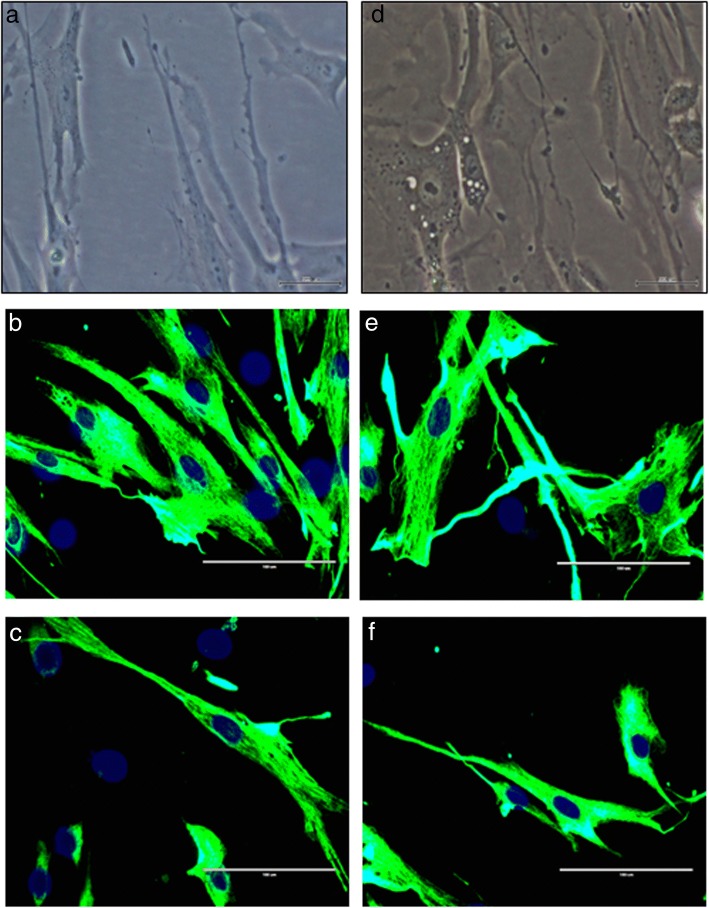
Fig. 1.
Morphology of young and senescent myoblasts for control and TRF-treated cells. Observation was carried out under phase contrast (a, d) and fluorescence microscopy (b, c, e, f) (× 40 magnification). The myoblast cells were stained with an antibody against desmin (green), and the nuclei were stained with Hoechst (blue). Control senescent myoblasts appeared larger and flatter with the presence of more prominent intermediate filaments (d, e) compared to control young myoblasts (a, b). Some of the TRF-treated senescent myoblasts (f) remained spindle-shaped which resembled young control while some exhibited flatter and larger morphology. No morphological changes was observed for TRF-treated young myoblasts (c)

