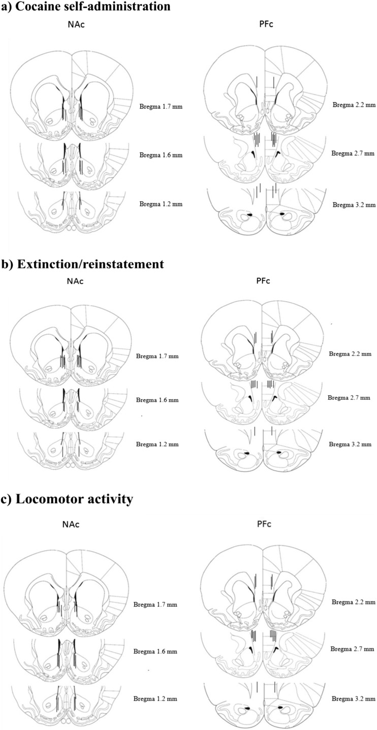
Fig. 2.
Histological verification of microinjection representative probe placements in the NAc (left panels) and the PFc (right panels) of rats that underwent cocaine self-administration (a), extinction/ reinstatement tests (b), and locomotor activity (c). Plates are taken from rat brain atlas Paxinos and Watson (1998) and the black line represent right placement of probes. Due to the large number of animals utilized for studies, bilateral placements are shown for only a subset of the experimental pool

