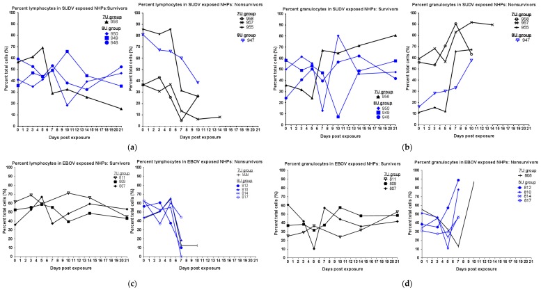
Figure 3.
Lymphocyte and granulocyte percentages in Macaca fascicularis exposed to low doses of Sudan virus (SUDV) or Ebola virus (EBOV). M. fascicularis were experimentally exposed to 0.01 plaque-forming units of either SUDV or EBOV with 7-uridine (7U) or 8-uridine (8U) genotype. During each scheduled sedation, and when possible at the time of death, blood specimens were collected and analyzed. Results of hematology analysis for all animals throughout the course of the study are shown. Normal values determined at the Texas Biomed clinical pathology lab—Lymphocytes: 43–77% and Granulocytes: 19–52%. NHPs—nonhuman primates. (a) Percentage of lymphocytes in SUDV-exposed NHPs. (b) Percentage of granulocytes in SUDV-exposed NHPs. (c) Percentage of lymphocytes in EBOV-exposed NHPs. No data are available for animal 811 on day 7 post-exposure. (d) Percentage of granulocytes in EBOV-exposed NHPs. No data are available for animal 811 on day 7 post-exposure.

