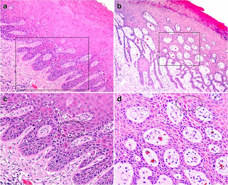
Fig. 1.
Example of differentiated VIN with characteristic features (HE stain), low magnification appearance (a, b), with corresponding higher magnification images (c, d). a A widened epithelium with parakeratosis and elongated rete ridges is seen. Nuclear atypia, premature keratinisation and cobblestone appearance are apparent (original magnification ×50). b Elongated rete ridges, a deep squamous eddy, nuclear atypia, and parakeratosis can be identified under low magnification (original magnification ×50). c Under higher magnification, macronucleoli can be seen. Angulated nuclei, individual cell keratinisation and cobblestone appearance (circled area) can be better appreciated (original magnification ×100). d Atypical cells with both open chromatin and hyperchromatic patterns are seen. There is cobblestone appearance (circled area) and individual cell keratinisation (original magnification ×100)

