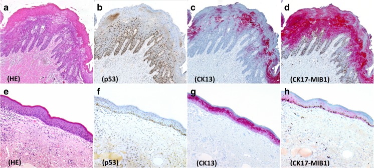
Fig. 3.
Immunohistochemistry in differentiated VIN (a–d) and Lichen sclerosus (e–h). a Differentiated VIN, HE stain b Overexpression of p53. c Weak, patchy CK13 staining. d Strong and diffuse CK17 expression, with increased MIB-1. e Lichen sclerosus, HE stain. f Wild-type p53 expression. g Diffuse staining of moderate intensity with CK13. h Very weak, patchy CK17 staining with increased MIB-1

