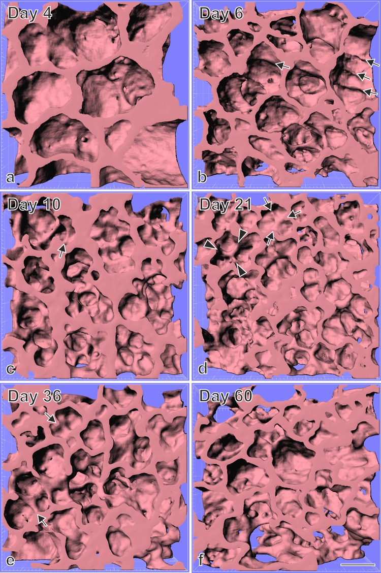
Fig. 4.
Visualization of alveolarization using synchrotron radiation based X-ray tomographic microscopy of rat lung parenchyma at postnatal days 4–60. Newly forming alveolar septa are recognized by low ridges which are upfolding from existing airspaces. As expected none of these ridges were observed in the larger saccules present during the saccular stage (postnatal day 4, a) but many after the start of alveolarization at days 6 and 10 (classical alveolarization, b, c, arrows). However, unexpectedly the same structures were observed at days 21 and 36 (d, e, arrows) which was taken as an indication that alveolarization continues at least until young adulthood. At day 60 the number of low ridges was below the limit of detection. Synchrotron radiation based X-ray tomographic microscopy was applied for 3D-imaging of lungs embedding for electron microscopy. Arrow head, mature septa; bar, 50 µm. Due to the perspective view the bar is only correct at the surface of the sample.
From Mund et al. (2008), by courtesy of Springer Nature Switzerland, Basel

