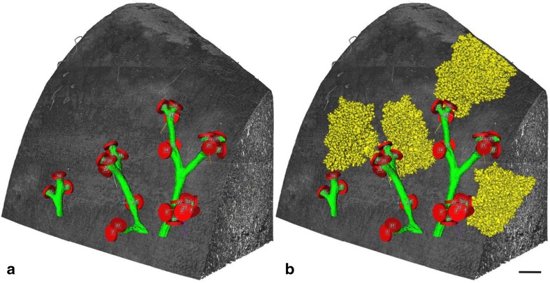
Fig. 8.
3D-visualization of acini branching off of distal conducting airways in a rat lung. Surface renderings of individual acini are shown in yellow. They are branching off of distal conducting airways (green) at the bronchioalveolar duct junction (labeled by red segmentation stoppers). The lung tissue is shown in shades of grey. a Conducting airways closed by segmentation stoppers at the acinar entrance. b Four non-neighboring acini are shown in addition of the structures shown in a. Bar 0.5 mm.
From Haberthur et al. (2013), by courtesy of Springer Nature Switzerland, Basel

