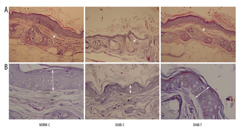
Figure 1.
Morphological change in the wound tissue of each group. Photomicrographs of (A) hematoxylin and eosin (H-E) staining at 400× and (B) Masson staining at 1000×. NORM-C rats showed re-epithelialization around the wound. The wound of the DIAB-C rats presented inflammatory cell infiltration. Re-epithelialization was observed in wounds of the DIAB-T rats. NORM-C – normal control; DIAB-C – diabetes control; DIAB-T – diabetes treated with the fusion protein.

