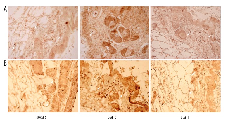
Figure 5.
Immunohistochemistry analysis of the granulation tissues (400×). (A) Staining of PDGF, PDGF-positive staining showed expression of proangiogenic factor; and (B) staining of CD34, CD34-positive staining was used to observe the small vessels in granulation tissues. NORM-C – normal control; DIAB-C – diabetes control; DIAB-T – diabetes treated with the fusion protein.

