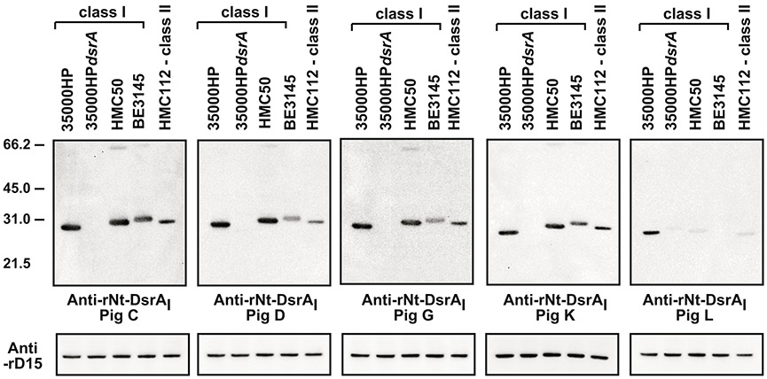
Figure 3. rNT-DsrAI antisera bind to homologous and heterologous denatured DsrA proteins.

Total cellular proteins (from approximately 1 × 10⁷ CFU) from the indicated strains were subjected to SDS-PAGE and Western blotting with the indicated polyclonal Abs at 1:10,000. All blots were incubated and developed concurrently. Blots were stripped and re-incubated with anti-rD15 [37] to show equal loading. Ponceau S staining and protein concentration were also used to determine equal loading in each lane (data not shown). Shown are representative blots from at least 2 independent experiments. The results presented in this figure reflect the reactivity of anti- rNT-DsrAI obtained one week after the 4th immunization, on the day of infection.
