10.
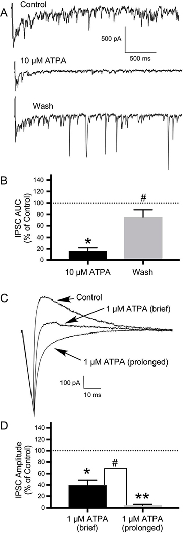
GluK1-containing KARs modulate inhibitory transmission in the OB. Reciprocal inhibition was induced by self-stimulation (using a brief depolarizing voltage pulse) of mitral cells and external tufted (ET) cells in OB slices to examine reciprocal inhibition from granule cells and periglomerular (PG) cells, respectively. A) With use of a CsCl electrode, this self-stimulation protocol evoked a flurry of IPSCs, which was quantified by determining the AUC. Compared with control, application of 10 μM ATPA significantly inhibited these reciprocal IPSCs, with significant recovery of IPSCs following a wash. B) Histogram showing the effect of 10 μM ATPA and recovery following a wash expressed as a percentage of the control IPSC AUC (N=5; *P ≤ 0.05 compared to control; # P≤ 0.05 compared to 10 μM ATPA). C) The effects of 1 μM ATPA on reciprocal inhibition in neurons in OB slices were also examined. With use of a KMeSO4 electrode, the self-stimulation protocol evoked a single IPSC. Both brief (30 seconds) and prolonged (5–10 minutes) application of ATPA significantly reduced IPSC amplitude, with greater effects with prolonged ATPA application. D) Histogram showing significant difference in the effects of brief (N=9) versus prolonged (N=7) duration of 1 μM ATPA application on IPSC amplitude expressed as a percentage of the control IPSC amplitude (#P ≤ 0.01; *P ≤ 0.01 compared to control; **P ≤ 0.01 compared to control). Values are the mean ± SEM.

