Figure 1:

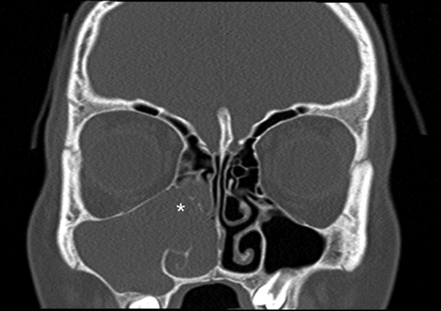
Coronal computed tomography (bone window) image showing a right-sided antrochoanal mass (white asterisk) extending in to the maxillary and ethmoidal sinuses.

Coronal computed tomography (bone window) image showing a right-sided antrochoanal mass (white asterisk) extending in to the maxillary and ethmoidal sinuses.