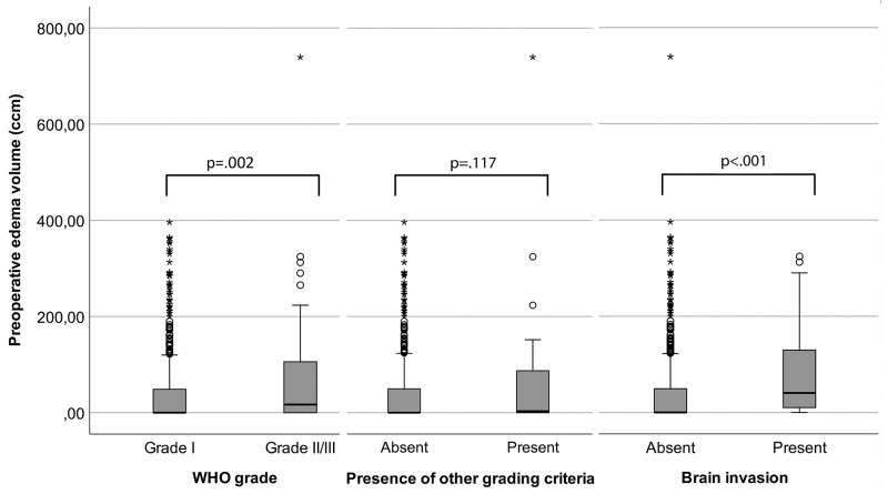
Figure 1. Boxpots visualizing the degree of association between peritumoural edema (PTBE) volume and histopathological findings.
High-grade histology was associated with increased PTBE volume (left, p=.002) and PTBE volumes were larger in invasive than in non-invasive meningiomas (p<.001, right). However, no association was found between edema volume and other histopathological grading criteria (p=.117). The boxes indicate upper and lower 25% quartile, the whiskers the minimum/maximum value within 1.5 IQR of the lower/upper quartile, the dots the outliers, the asterisks the extreme values, and the heavy horizontal line indicates the median (ccm=cubic centimeter, *high-grade=grade II and III meningiomas.).

