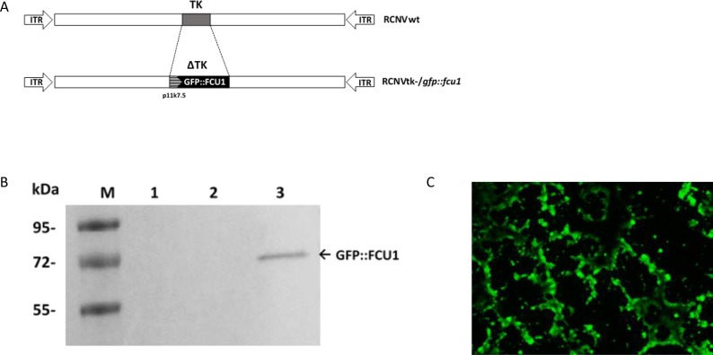
Figure 2. Generation of RCNV expressing the GFP::FCU1 fusion gene and evaluation of the GFP::FCU1 protein expression.
(A) Schematic representation of Raccoonpox viruses used in this study. RCNtk-/gfp::fcu1 contains a deletion of TK gene and insertion of a fusion gene between eGFP and FCU1 genes. The GFP::FCU1 fusion gene is driven by the synthetic p11k7.5 promoter. (B) Western blot detection of the GFP::FCU1 protein by anti-FCU1 monoclonal antibody. Lane 1 (left to the right), mock-infected LoVo cells; lane 2, LoVo cells infected with RCNVwt; lane 3, LoVo cells infected with RCNtk-/gfp::fcu1. Molecular weight standards are shown in kDa on the left. The presence of GFP::FCU1 fusion protein (Mr 72,000) is indicated (arrow). (C) Fluorescent microscopy showing the GFP::FCU1 protein expression. HCT 116 cells were infected with RCNtk-/gfp::fcu1 at MOI 0.001 and transgene expression (GFP) was monitored 72 h post infection by fluorescent microscopy.

