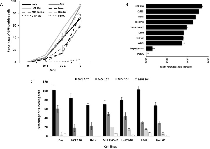
Figure 4. Infection, replication and oncolytic activity of RCNtk-/gfp::fcu1.
(A) Infection susceptibility of human cells to RCNtk-/gfp::fcu1. Cells were infected at the indicated MOI with RCNtk-/gfp::fcu1 and the percentage of GFP-positive cells was determined by flow cytometry at 16 h post infection. The results were obtained from three separated experiments ± SD. (B) Replication in tumor cell lines and in primary human cell. Cells were infected at MOI 10–3, except PBMC at MOI 10–1, and harvested after 3 days of infection. Results are expressed as viral fold amplification and were obtained from three separated experiments ± SD. Asterisk denotes absence of amplification. (C) Oncolytic activities of RCNtk-/gfp::fcu1 by measuring the cell viability 5 days after infection of different cancer cell lines. Tumor cells were infected at a MOI ranging from 10–4 to 10–1 and cell viability was determined by ViCell cell counter automate based on trypan blue exclusion method. Each data represents the mean of triplicate determinations ± SD.

