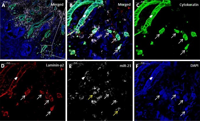
Fig. 3.
Colon cancer specimen with miR-21 positive tumor budding cells. a Merged image of the tumor periphery, cytokeratin (green), miR-21 (white), laminin-5γ2 (red) and DAPI (blue). b Magnification of an area containing tumor budding cells (arrows) and a malignant glandular structure (arrowhead). c Single channel image for cytokeratin. d Single channel image of laminin-5γ2. The adenocarcinoma cells show laminin-5γ2 overexpression. e MiR-21 expression is only seen in a few tumor buds (yellow arrows) and focally in the malignant gland structure (arrowhead). f DAPI image

