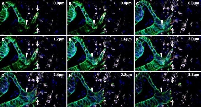
Fig. 4.
Tumor cell budding in confocal stack of images. Example of a confocal stack of images covering 3.2 µm in the z-axis in the tissue section, acquired from a digital whole slide of an adenocarcinoma tissue section stained for miR-21 (white), cytokeratin (green) and laminin-5γ2 (red). At baseline (0.0 µm) a budding cancer cell event (white arrow) and a malignant gland structure is noted. The stack reveals direct connection between the gland and budding cell event in the stack at 2.4–2.8 µm, identifying the tumor budding cells as a ‘branching event’

