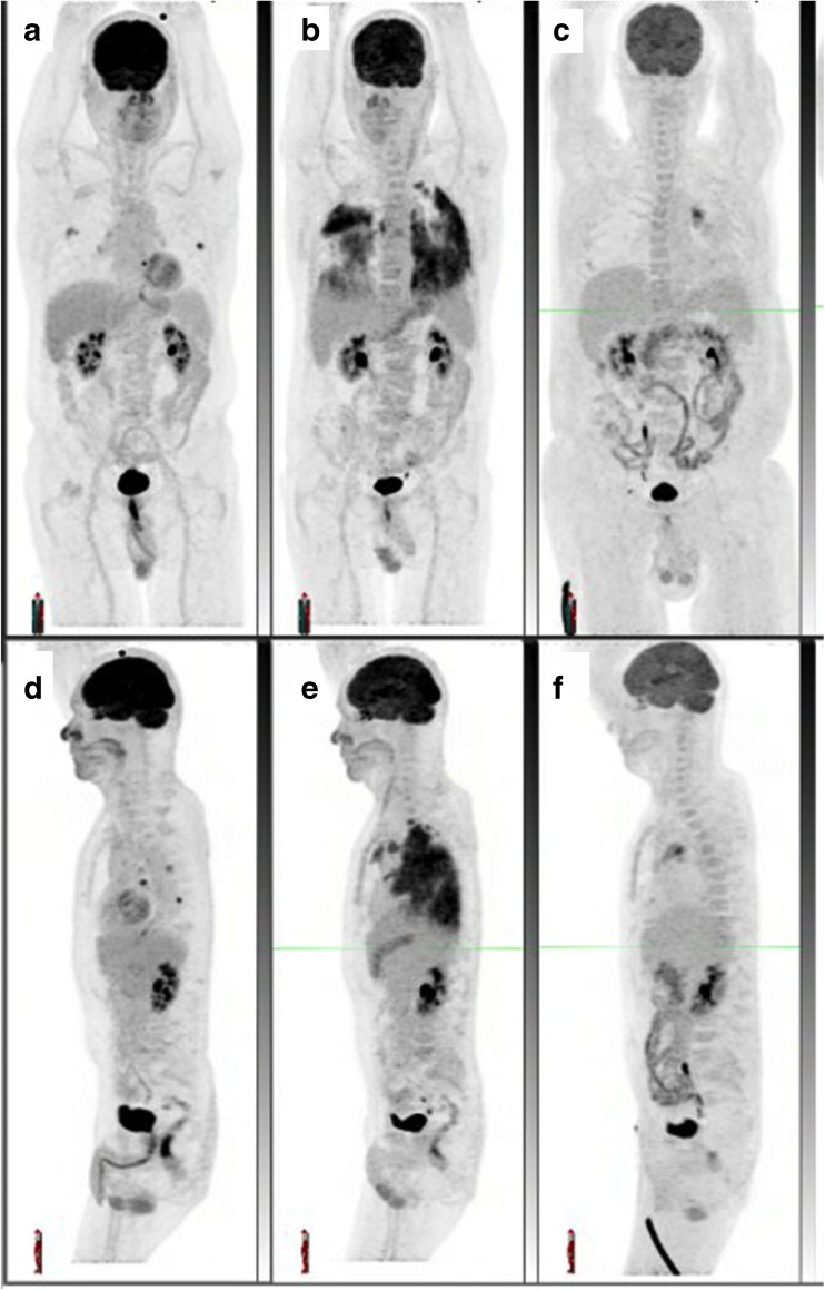
Fig. 4.
Serial maximum intensity projection images (a–c anterior, (d–f ) left lateral) show the development and resolution of pneumonitis. Note the dominance of parenchymal changes in the dependent lung, which is typical. There was a complete metabolic response with low-grade left hilar changes (c, f) consistent with reactive lymphadenopathy

