Fig. 1.
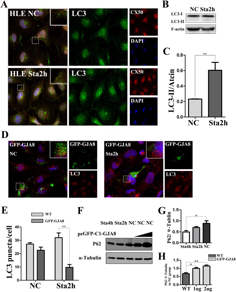
Overexpressed GJA8 protein modulated the level of LC3 and P62 in HLE cells. a HLE cells stained for GJA8 (red) and LC3 (green) in the NC and Sta2h groups. b Western blot with LC3-I/II in NC and Sta2h groups in HLE cells. c The average level of LC3-II/Actin of three independent experiments. d Transfected with peGFP-C1-GJA8 in the NC and Sta2h groups, HLE cells stained for LC3 (red). e Mean number of LC3 puncta for each treatment (n = 3 wells, 3 independent experiments, > 50 cells per experiment). f HLE cells for corresponding treatment and transfected with peGFP-C1-GJA8 for 0ug or 1ug or 2ug, testing the expression of P62 and α-Tubulin. g The average level of P62/α-Tubulin of three independent experiments in Sta4h/Sta2h/NC group. h The average level of P62/α-Tubulin of three independent experiments transfected with peGFP-C1-GJA8 for 0ug or 1ug or 2ug in NC group. All values are represented as the mean + SEM; *p < 0.05, **p < 0.01 indicate significant differences with corresponding groups. Nuclei are stained with DAPI

