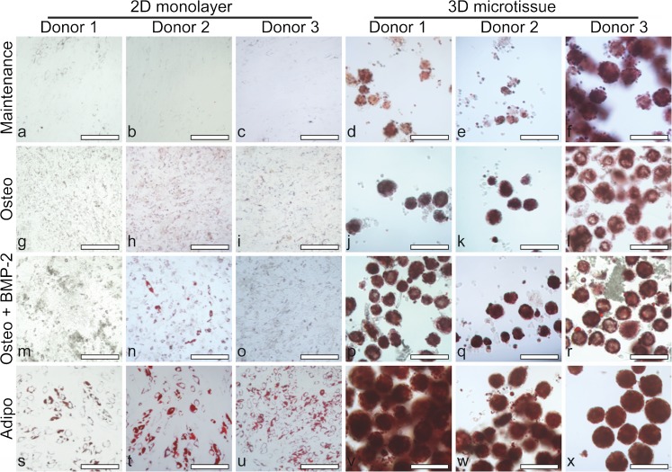
Fig. 8.
Bright field microscopy to visualise lipid vacuoles (red) by Oil Red O staining in 2D cultures for BMSC donor 1 (a, g, m and s), donor 2 (b, h, n and t) and donor 3 (c, i, o and u) and in 3D cultures for BMSC donor 1 (d, j, p and v), donor 2 (e, k, q and w) and donor 3 (f, l, r and x). Day 21 cultures are shown following growth in maintenance medium (DMEM + 10% FBS) (a–f), osteogenic medium (Osteo) (g–l), osteogenic medium + BMP-2 (Osteo + BMP-2) (m–r) or adipogenic medium (Adipo) (s–x). Scale bars = 200 μm

