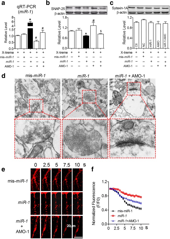
Fig. 4.
MiR-1 inhibited SNAP-25 protein expression and presynaptic vesicle release. a Verification of miR-1 in NRNs after transfection. PLevene = 0.001, One-way ANOVA: F = 41.566, P < 0.0001; Fisher’s PLSD test: Pmis-miR-1: miR-1 < 0.0001, PmiR-1: AMO-1 < 0.0001. n = 3 batches of cells for each group. b Expression of SNAP-25 protein in NRNs was downregulated by miR-1 determined by western blot analysis. PLevene = 0.32, One-way ANOVA: F = 4.667, P = 0.008; Fisher’s PLSD test: Pmis-miR-1: miR-1 = 0.04, PmiR-1: AMO-1 = 0.001. n = 5 batches of cells for each group. c The expression of syntaxin-1A protein in NRNs was not influenced by miR-1. PLevene = 0.151, One-way ANOVA: F = 1.033, P = 0.422. d Morphologic change of the transfected primary cultured neurons. e Representative FM1–43 fluorescent signalling changes in each group after 70 mM KCl stimulation. f Co-transfection of AMO-1 with miR-1 improved the decline in FM1–43 signalling decline (F/F0) in NRNs compared with transfection of miR-1 alone. χ2Mauchly = 1652.465, P < 0.0001; Ftotal (19, 1407) = 48.263, P < 0.0001; Fisher’s PLSD test: Pmis-miR-1: miR-1 < 0.0001, PmiR-1: miR-1 + AMO-1 < 0.0001, Pmis-miR-1: miR-1 + AMO-1 < 0.0001. n = 25 neurons from 3 batches of cells for each group

