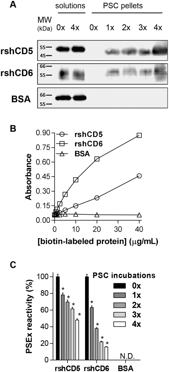
Fig 1. CD5 and CD6 ectodomains bind PSC tegumental antigens.

(A) Biotin-labeled rshCD5, rshCD6 and BSA protein solutions (20 μg/mL) were sequentially incubated (4 times) with PSC suspensions (5,000 PSC; viability ≥90%), and pellets and solutions run on SDS-PAGE and further Western blotted with HRP-streptavidin. Lanes 1 and 2: protein solutions before (0x) and after 4 sequential incubations with PSC (4x), respectively. Lanes 3 to 7: PSC pellets after 0x to 4x sequential incubations. (B) ELISA assays showing the binding of increasing amounts of biotinylated rshCD5, rshCD6 and BSA proteins to PSEx-coated plates. (C) ELISA assays showing the binding of supernatants from sequential PSC incubations of biotin-labeled rshCD5, rshCD6 and BSA depicted in (A), to PSEx-coated plates. N.D. not detected. (*) Significant differences (Student’s t-test, P <0.05) respect to 0x results.
