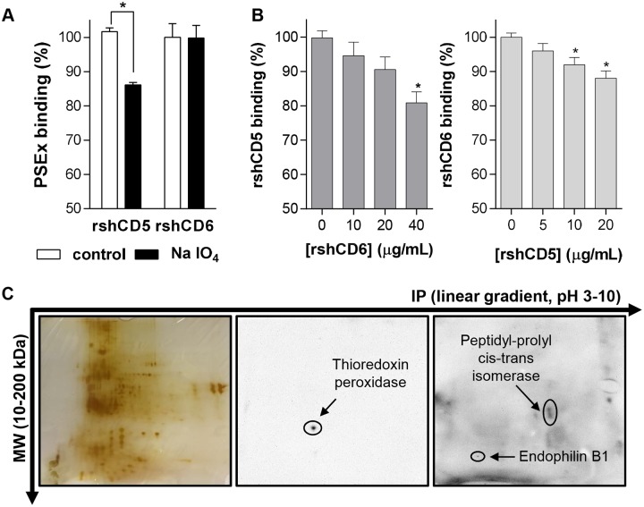
Fig 2. Tegumental antigens recognized by rshCD5 and rshCD6 are different.
(A) ELISA assays showing binding of rshCD5 and rshCD6 to PSEx-coated plates undergoing metaperiodate treatment (NaIO4) or not (control). Binding to metaperiodate-resistant antigens was assessed as the percentage of absorbance values in treated wells respect to untreated wells (triplicates in each case). (B) Competition binding assays in which binding of a fixed amount of biotinylated rshCD5 (20 μg/mL; left) or rshCD6 (10 μg/mL; right) to PSEx-coated ELISA were competed with increasing amounts of unlabeled rshCD6 and rshCD5 proteins, respectively. Ligand overlapping was assessed (in triplicates) as the percentage of absorbance values in competed wells respect to non-competed wells (0 μg/mL of unlabeled protein). (C) 2D SDS-PAGE resolved PSEx fractions were either silver stained (left) or electro-transferred onto PVDF membranes for further Western blotting with biotin-labeled rshCD5 (middle) or rshCD6 (right). (*) Significant differences (Student’s t-test, P <0.05) respect to control (A) or uncompleted (B) wells.

