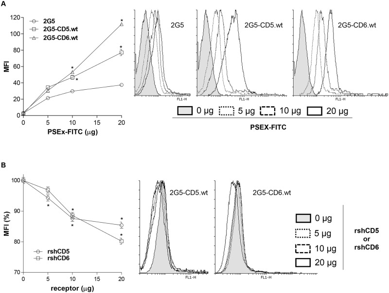
Fig 3. Native membrane-bound CD5 and CD6 receptors retain PSEx-binding capacity.
(A) Flow cytometry analyses of 2G5, 2G5-CD5.wt or 2G5-CD6.wt cells stained with increasing amounts of FITC-labeled PSEx. Represented are the mean fluorescence intensity (MFI) values (left) and a representative flow cytometry histogram from each case (right). (B) Competition binding experiments in which 2G5-CD5.wt or 2G5-CD6.wt cells were stained with a fixed suboptimal amount of FITC-labeled PSEx in the presence or absence of different amounts of unlabeled rshCD5 or rshCD6 proteins. Both experiments were performed in quadruplicates and results are shown as mean +/- SD. (*) Significant differences (Student’s t-test, P <0.05) respect to 2G5 cells (A) or cells with 0 μg of competing proteins (B).

