Figure 5.
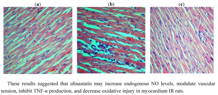
Histopathological changes in the myocardium at three groups. (a) The sham group, with a well-arranged cardiac cells and integrated membrane. (b) The IR group, showing swelling myocardial cells, a disordered striated cardiac muscle, and local myocardial necrosis. A great number of erythrocytes are present and local infiltration of inflammatory cells is observed. (c) The IR + ulinastatin group, showing a well-arranged cardiac cells and a tiny amount of neutrophil infiltration.

