
Figure 4. Inhibition of TBK1/IKKε leads to TNF-induced RIPK1 activation and increased complex II formation.
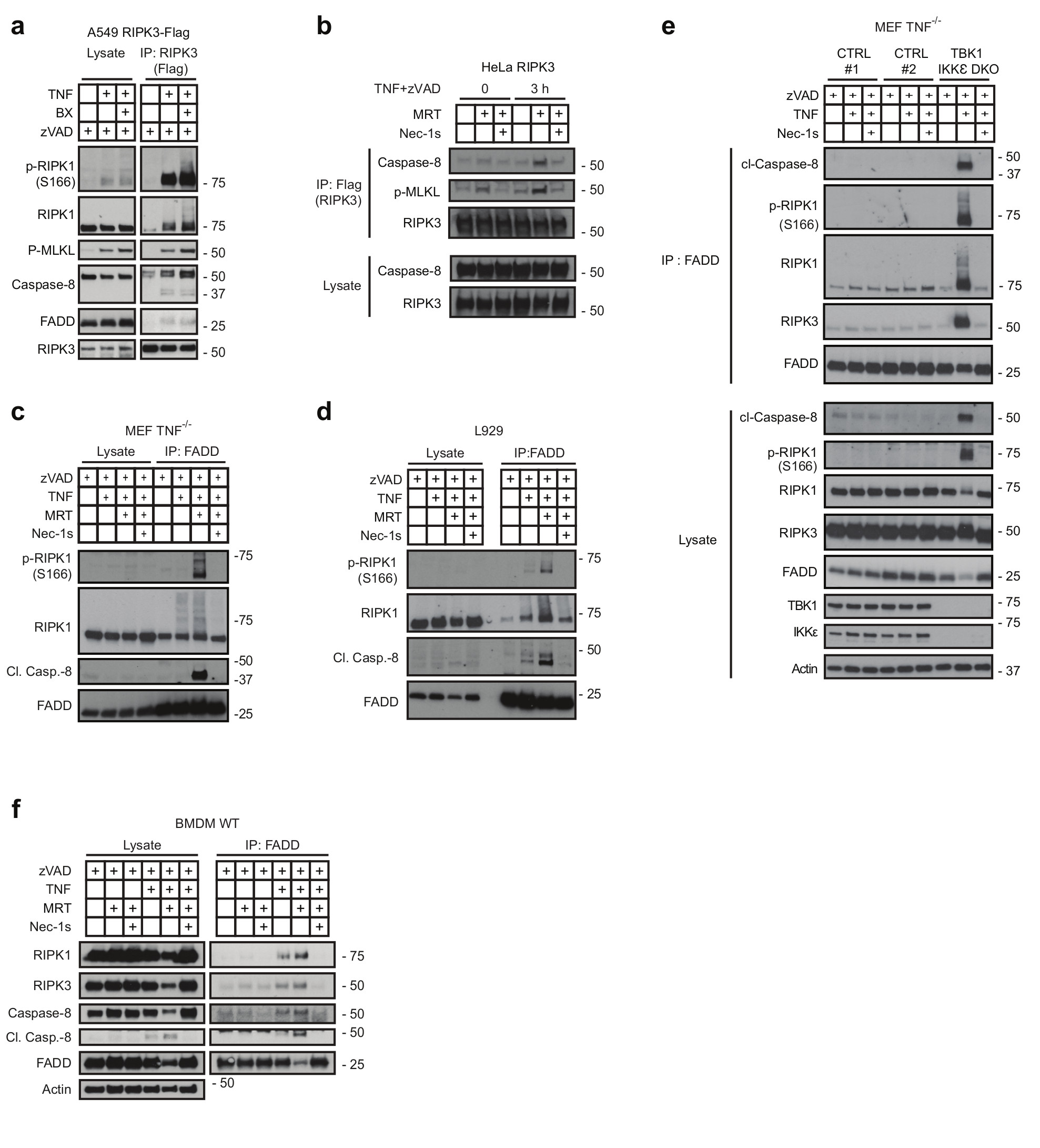
(a) A549 and (b) HeLa cells both overexpressing FLAG-tagged RIPK3 were treated with or without BX-795 and zVAD (a) or MRT, zVAD and Nec-1s (b) and were stimulated with TNF (500 ng/mL) for 3 hours. Complex II was then FLAG-immunoprecipitated and analysed by western blot. (c-e) MEFs (c), L929 (d), MEF TNF-/- ;TBK1/IKKε DKO cells and corresponding MEF TNF-/- control cells (e) and primary BMDMs (f) were pre-treated with MRT in combination with zVAD and Nec-1s as indicated and stimulated with TNF (500 ng/mL), for either 6 hours (c, e, f) or 4 hours (d), respectively. Complex II was then immunoprecipitated with a FADD antibody and analysed by western blot. (a-f) One representative experiment is shown out of two independent experiments. Unprocessed original scans of blots are shown in Supplementary Figure 7.