
Figure 1. Cytoarchitecture of the meninges, brain vasculature and pathways of paravascular recirculation.
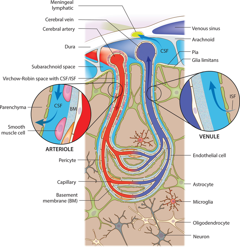
A schematic representation of the brain meninges constituted by dura, arachnoid and pia layers. Lymphatic vessels that are present in the meningeal dura drain components of the cerebrospinal fluid (CSF) that fills the subarachnoid space. Arising from the brain surface, cerebral arteries extend into pial and then subpial arteries. Higher caliber pial arteries extend into smaller caliber arterioles (both wrapped by smooth muscle cells) that dive into the brain parenchyma. Clearly defined paravascular spaces of about 50–100 nm, the Virchow-Robin spaces, are filled with CSF that flows into deeper brain regions, along the arterioles and capillaries, and diffuses through the glia limitans into the parenchyma. Efflux of interstitial fluid (ISF) happens through paravenous spaces back into the subarachnoid CSF.