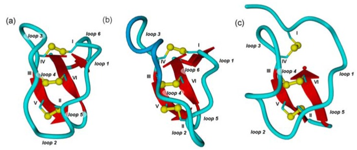
Figure 1.
Cartoon diagrams of prototypical cystine knots. Loops are depicted in light blue and numbered according to their appearance in the sequence, α-helices in dark blue, β-sheets in red, and cysteines in yellow with Roman numerals according to their appearance in the sequence. (a) Möbius cyclotide kalata B1. (PDB-ID: 1NB1) (b) Bracelet cyclotide cycloviolacin O2. (PDB-ID: 2KNM) (c) Acyclic inhibitor cystine knot ocMCoTI-II (PDB-ID: 2IT8). Structures modeled with Yasara Ver. 12.4.1.

