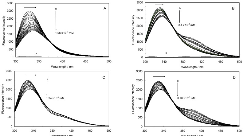
Figure 1.
(A) Fluorescence emission spectra of the hTf-ROP system. [hTf] = 3.8 × 10⁻³ mM and [ROP] was increased from 0 to 1.06 × 10⁻⁴ mM; (B) Fluorescence emission spectra of the hTf-ASA system. [hTf] = 3.8 × 10⁻³ mM and [ASA] was increased from 0 to 8.4 × 10⁻³ mM; (C) Fluorescence emission spectra of the (hTf-ASA)-ROP system. [hTf] = 3.8 × 10⁻³ mM and [ROP] was increased from 0 to 1.24 × 10⁻⁴ mM, [ASA] = 4.62 × 10⁻³ mM; (D) Fluorescence emission spectra of the (hTf-ROP)-ASA system. [hTf] = 3.8 × 10⁻³ mM and [ASA] was increased from 0 to 8.28 × 10⁻³ mM, [ROP] = 1.33 × 10⁻⁵ mM. The a and b represent the curves of ROP and ASA, respectively. All experiments were performed under identical conditions (T = 298 K; pH 7.4, λex = 280 nm). The horizontal arrow shows the red shift.

