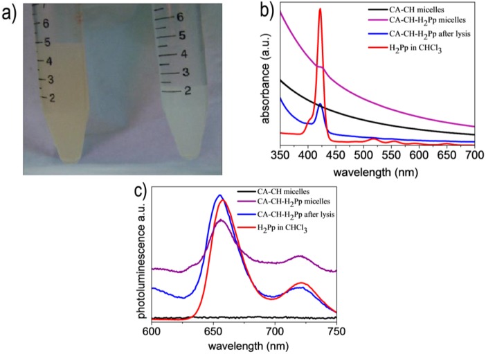
Figure 3.
(a) Comparison of a CA-CH-H2Pp (left side) micellar nanodispersion in buffer solution with a CA-CH (right side) blank sample. (b) UV–visible absorption and (c) photoluminescence spectra respectively of CA-CH micelles in buffer solution (black line), CA-CH-H2Pp micelles in buffer solution (purple line), CA-CH-H2Pp micelles after their lysis and solubilisation in chloroform (blue line) and H2Pp hybrid solution in chloroform (red line).

