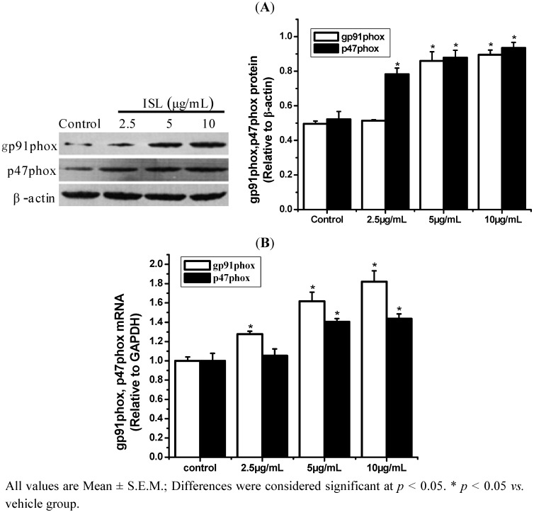
Figure 5.
The effect of ISL on p47phox and gp91phox expression in HL-60 cells. (A) Cells were treated with ISL (0, 2.5, 5, or 10 μg/mL) for 72 h, and protein was extracted rapidly for Western blot analysis. β-Actin was used as the internal control. (B) The levels of p47phox and gp91phox mRNA expression in ISL-treated HL-60 cells. GAPDH was used as an internal control.

