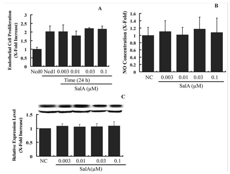
Figure 6.
Sal A does not constrain endothelial cell proliferation and does not influence eNOS expression. Confluent HUVECs (starved for 24 h in FCS-free DMEM) were treated with SalA at different concentrations (0.003–0.1 μM) for 24 h. Crystal violet staining was used to detect cell proliferating activity (A); Supernatants were then collected and NO levels were measured by using the Griess reagent. Concentration was adjusted according to cell number (B); Western blot analysis was performed to detect the relative expression level of eNOS in HUVECs. GAPDH was used for normalization (C). The relative level of the cell number and NO concentration is expressed as x-fold increase compared to that of the normal control group. Data shown are the mean ± SEM from three independent experiments.

