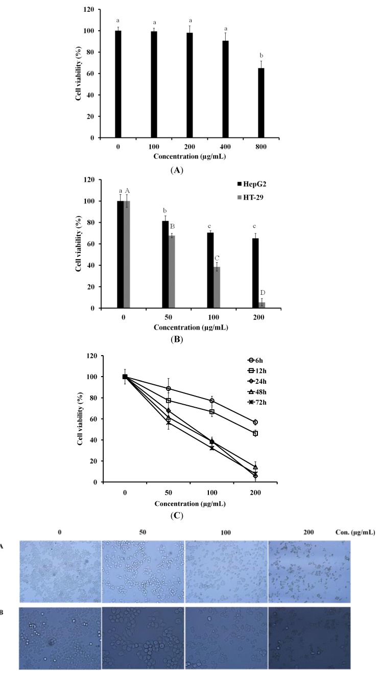
Figure 1.
Cytotoxicity and anti-proliferative activity of Luffa echinata. (A) The CCD-986Sk cells were plated in 96-well plates and then exposed to 100, 200, 400, and 800 µg/mL of L. echinata extract for 24 h. With in a column, values with the same superscript letters are not significantly different from each other at p < 0.05; (B) HepG2 and HT-29 cells were plated in 96-well plates and then exposed to 50, 100, and 200 µg/mL of L. echinata extract for 24 h. The cell viability of HT-29 cells treated by L. echinata extract was measured using an MTT assay. a–c indicated the statistical significance of the anti-proliferative activity of various concentrations of LER extract on HepG2 cells (p < 0.05). A–D indicated the statistical significance on HT-29 (p < 0.05); (C) HT-29 cells were incubated with 50, 100, and 200 µg/mL of L. echinata extract for 6, 12, 24, 48, 72 h, respectively. The cell viability of HT-29 cells treated by L. echinata extract was measured by MTT assay. (D) Cell culture morphology was assessed by light microscopy after incubation with various concentrations of L. echinata extract for 24 h.

