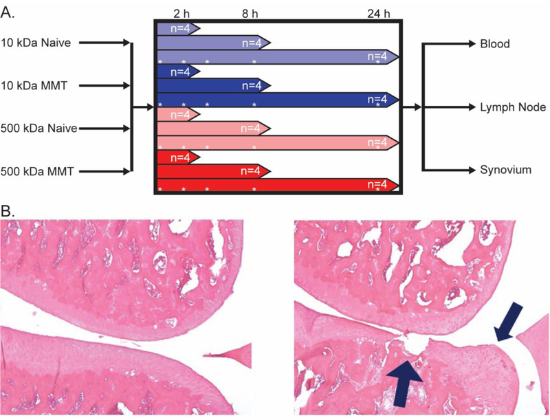
Figure 1:

A. Study design, animals were grouped by sacrifice point post-injection. At time of sacrifice, plasma as well as synovial and lymph tissue was collected. Asterisks represent in vivo imaging timepoints B. Histology of sham (Left) and MMT-operated (Right) rats 21 days post-surgery. Frontal sections of the medial aspect of rat knee joints were stained with Toluidine Blue. Sham exhibits normal cartilage, subchondral bone and synovium. MMT surgery. Prominent chondrophyte/osteophyte present adjacent to severe cartilage erosion. Chondrocyte loss is complete to the deep zone and significant proteoglycan loss can be detected all the way to the tidemark.
