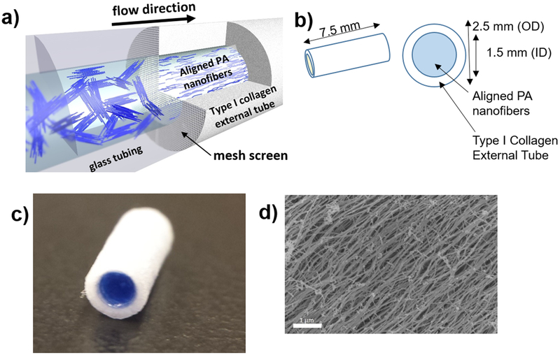
Figure 1.
Aligned nanofiber neurografts were thus constructed: (a) a solution of peptide amphiphile nanofibers is flowed across a mesh screen into the type I collagen external tube, which is then submerged into 25mMol calcium chloride solution to induce gelation, (b) neurograft schematic, OD (outer diameter), ID (inner diameter), (c) macro-photograph of PA nanofiber neurograft, PA nanofiber solution dyed blue for contrast within type I collagen external tube. d) Scanning electron microscopic image of aligned nanofibers within the neurograft, scale bar 1 μm.

