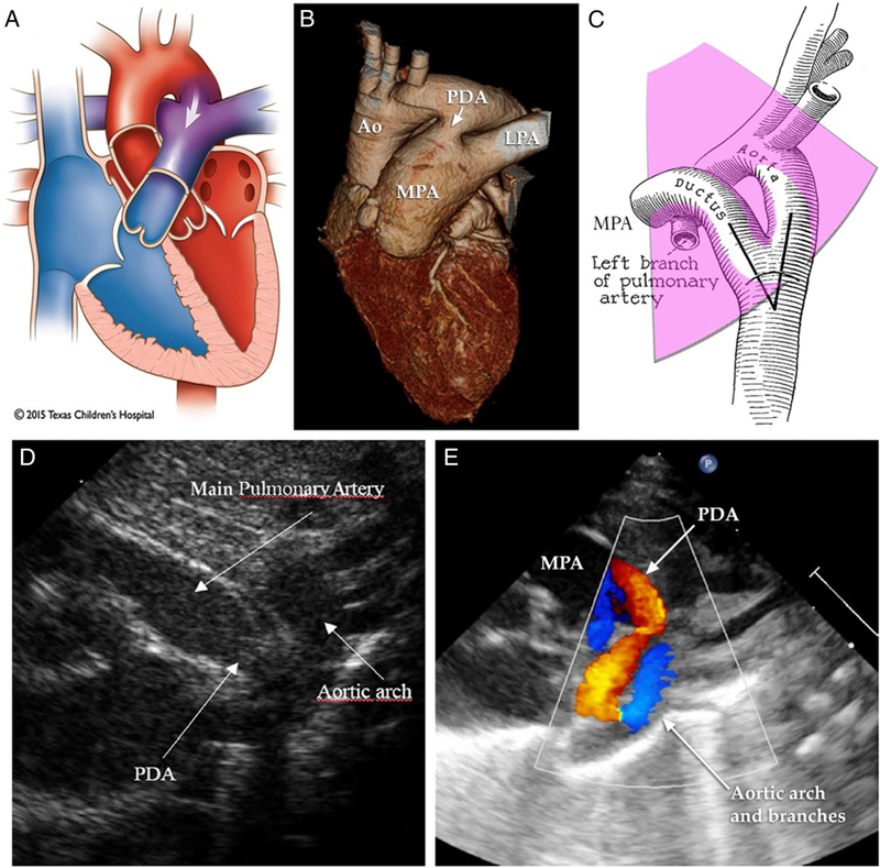
Figure.
Evaluation of patent ductus arteriosus (PDA) with echocardiography. A, B. Left-to-right (L-R) shunt (indicated by arrows) via the ductus produces most of the physical signs and complications related to PDA. C–E.Short-axis andsuprasternal views reveal structural relationships and color Doppler flow patterns indicative of L-R shunt across the ductus arteriosus. Ao=aorta; LPA=left pulmonary artery; MPA=main pulmonary artery. Images adapted or reprinted with permission from Texas Children΄s Hospital,61)(62) and by Creative Commons license.(63)

