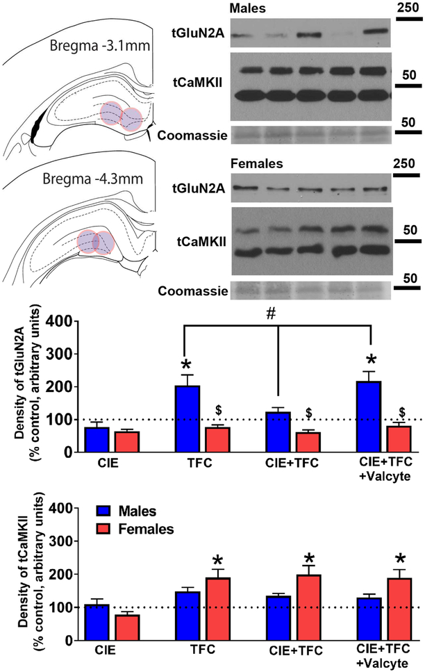
Figure 5:
GluN2A and CaMKII expression is distinctly effected by TFC and CIE in male and female rats. (a) Schematic showing location of tissue punches taken in the dorsal DG of the hippocampus. (b-c) Representative Western blots for protein expression in male (b) and female (c) DG enriched tissue. Lane 1- Naïve control; 2- CIE; 3- TFC; 4- CIE+TFC; 5- CIE+TFC+Valcyte. Molecular weight of ladder in kDa is indicated to the right of the blots. (d-e) Density of protein expression for total GluN2A and CaMKII in dorsal DG from male and female rats. #p<0.05 indicating significant interactions; *p<0.05 compared to drug and sucrose naïve age matched controls; &p<0.05 vs. CIE+TFC-Valcyte; $p<0.05 vs. males by three-way ANOVA followed by posthoc tests. Data shown are represented as mean +/− SEM. n = 6 behavior naïve males, n = 9 CIE males, n = 16 CIE naïve TFC males, n = 11 Valcyte− CIE TFC males, n = 14 Valcyte+ CIE TFC males; n = 6 behavior naïve females, n = 9 CIE females n = 13 CIE naïve TFC females, n = 15 Valcyte− CIE TFC females, n = 10 Valcyte+ CIE TFC females.

