Figure 4.

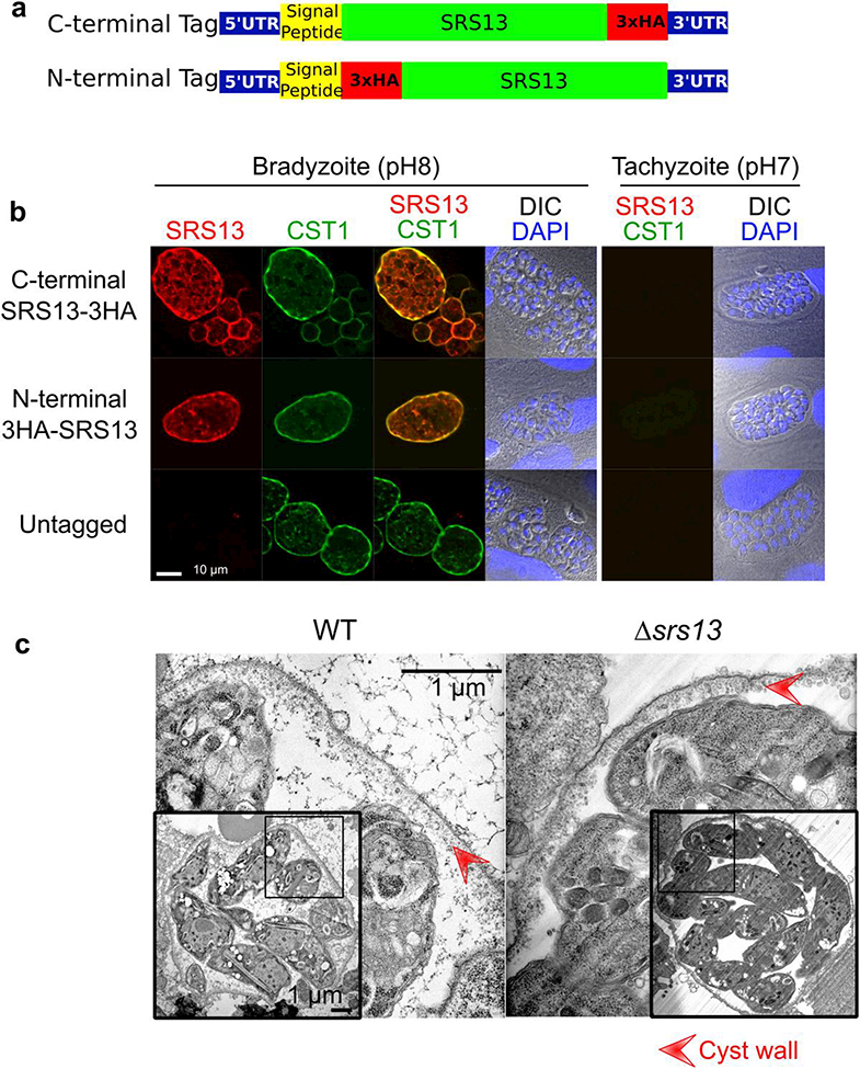
a. Schematic for the HA tagging strategies. For C-terminal tagging a 3xHA sequence was added immediately before the stop codon. For N-terminal tagging a 3xHA sequence was added immediately after the predicted signal peptide sequence..
b. SRS13 is localized to cyst wall and bradyzoite matrix but not in tachyzoite. Immunocytochemical image of in vitro T. gondii cultured in either bradyzoite condition (pH8 for 5 days) or tachyzoite condition (pH7 for 3 days) probed with anti-HA antibody (red), rabbit polyclonal anti-CST1 antibody (green), and DAPI (blue).
c. SRS13 is not required for the formation of in vivo cyst wall. Transmission electron (TEM) micrograph of in vivo mouse brain cysts in low and high magnifications are shown. The lower magnification TEM image of whole cysts are in the insert panels. Red arrowheads indicate the cyst wall.
