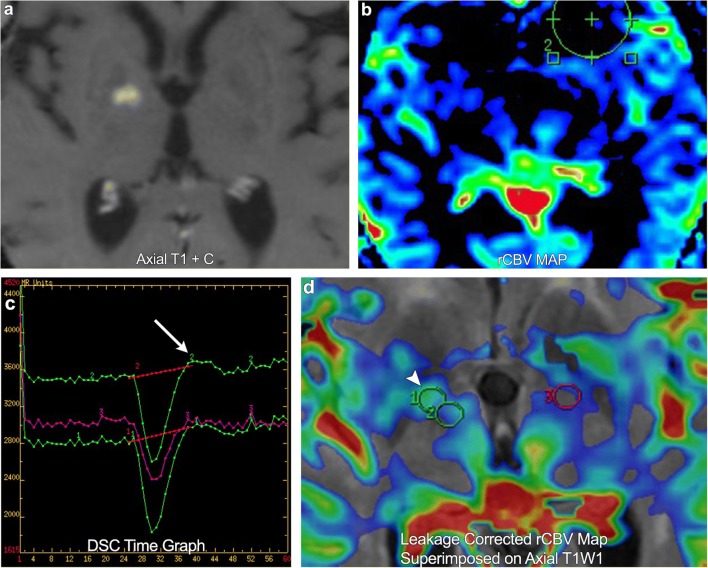
Fig. 5.
A 73-year-old female with right frontal lobe glioblastoma multiforme. Post-treatment imaging demonstrates a new region of enhancement in the right basal ganglia. DSC rCBV map shows no definite increased perfusion. However, the time graph demonstrates overshoot (arrow). DSC leakage-corrected map superimposed on axial T1WI now shows asymmetric elevated rCBV in the enhancing lesion (arrowhead), compatible with a new site of tumour

